
Cardiac MRI in Baroda
Advanced cardiac MRI for tissue characterization, morphology & flow at Baroda Imaging Centre — Alkapuri, Raopura & Old Padra Road.
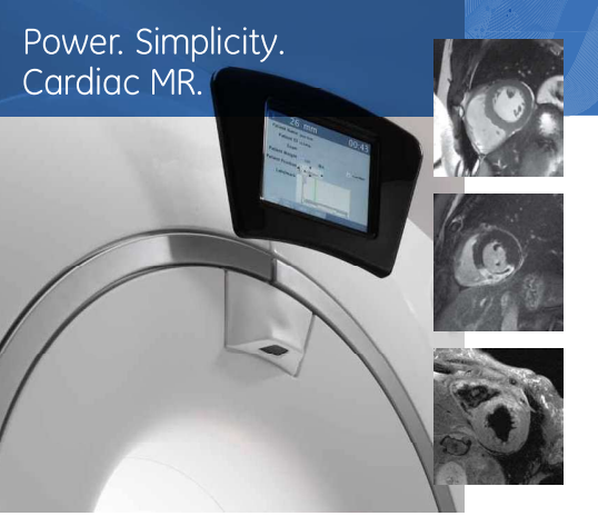
Cardiac MRI
Tissue characterization and function.
Morphology and flow.
Cardiac MR from GE Healthcare combines power and simplicity, to help you see well beyond just cardiac anatomy. It gives you comprehensive clinical information on hard-to-detect cardiac conditions that no other single imaging modality can provide.
Answer Beyond Anatomy
GE Cardiac MR gives you the power to effectively characterize myocardial tissue, so you can assess a wide range of cardiac diseases, and iron overload other modalities can't see. With GE Cardiac MR you have the ability to visualize not just anatomy, but deep inside the myocardium to see exactly what's happening. MR also has the power to examine patients easily and effectively — from pediatric patients to those concerned about radiation exposure, to women with silent ischemia. With GE Cardiac MR, you see things you can't see any other way — for your shortest path to diagnostic confidence.
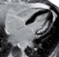
Viability Imaging
T-2 Weighted edema imaging
T2* Imaging
From ischemic heart disease and cardiomyopathies, to congenital heart disease and MR angiography, GE Cardiac MR gives you quick, easy ways to assess a wide range of cardiac disease, free of ionizing radiations.
See It All.
MR
Gated 2D FIESTA / FGRE Cine
High-quality cardiac images for wall motion assessment
Optimized for cardiac imaging, the 2D FIESTA/FGRE Cine pulse sequence provides excellent blood-to-myocardium contrast. Short TR/TE allows for the acquisition of high-quality cardiac images that are less sensitive to turbulent blood flow and off-resonance artifact.
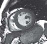
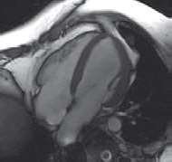
2D FIESTA Cine from 1.5T (left) & 3.0T (right)
MRI Echo
Image the heart in real time — without gating.
Excellent for imaging cardiac patients who have arrhythmias or difficulty holding their breath, GE MR Echo generates excellent real-time images without the need for ECG gating or breath-holds. Interactive user-interface allows you to identify primary cardiac planes with ease.
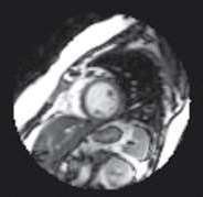
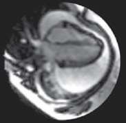
MR Echo routinely produces gated free-breathing images like these
Time Course (FGRE TC / FGRE-ET / FIESTA TC)
Perform fast, robust cardiac time-course studies with clinical confidence.
This is the first choice for stress studies to differentiate between stunned versus infarcted myocardium. FGRE TC delivers excellent temporal and spatial resolution and T1 contrast to aid in more confident diagnosis. The Fast Gradient Echo Time Course displays a superb contrast-to-noise ratio and is less sensitive to off-resonance and eddy current effects.
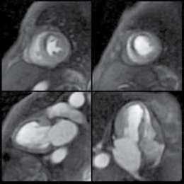
Multi-plane FGRE Time Course showing myocardial infarction in both short-axis and long-axis views.
2D / 3D Myocardial Delayed Enhancement (MDE)
Determine myocardial tissue viability simply and reliably.
Performed in a single breath-hold, 2D/3D MDE provides fast, simple, and reliable assessment of myocardial tissue viability or fibrosis. It works by improving the contrast-to-noise ratio between infarcted and normal myocardium.
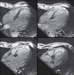
High-resolution free-breathing 3D MDE enables flexible reformat into any desired plane.
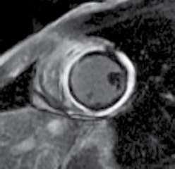
2D MDE Showing Myocarditis with LV pericardium involvement.
Cine IR
Optimal TI time, every time.
A simple, fast, and easy-to-use multiphase FGRE-Cine acquisition performed in a single breath-hold. Cine IR captures image contrast evolution at different TI (Inversion Time) times to quickly determine the optimal TI for myocardial delayed enhancement (MDE). It is also used to detect amyloid, vasculitis, and other tissue abnormalities through the TI evolution.
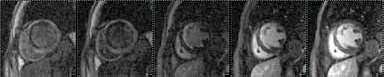
Cine IR showing TI evolution of normal and infarcted myocardium.
T2*
Measure cardiac iron levels noninvasively.
T2* is a useful technique for evaluating cardiac iron overload. It enables iron assessment in the cardiac tissue so that chelation treatment can be adjusted appropriately to help prevent complications. MRI is the only noninvasive modality in clinical use capable of detecting cardiac iron deposits.
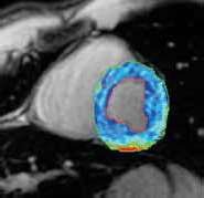
T2* assessment for iron overload.
T2w Blackblood Imaging
Identify inflammation, edema, and visualize masses and valves.
This double and triple IR fast-spin-echo (FSE) technique provides T1w or T2w images with excellent blood suppression. This allows for clear visualization of edema, cardiac masses, or valve leaflets.
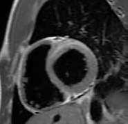
Edema with T2 elevation observed in a patient with acute myocarditis.
3D Heart Coronary Artery Imaging with Cardiac Navigator
See whole-heart morphology in clear detail.
This free-breathing, navigator-assisted technique shows you the entire heart in exquisite anatomical detail. With 3D Heart, you can image the coronary arteries, evaluate vascular structures, and assess congenital heart disease (CHD) noninvasively in adult and pediatric patients without sedation, anesthesia, radiation, contrast media, or the limitations of ultrasound. Navigator echo tracks the diaphragm motion to synchronize the acquisition to the patient's end-expiration respiratory phase, enabling free-breathing acquisition. Real-time motion correction further minimizes respiratory ghosting artifacts while improving the scanning efficiency.
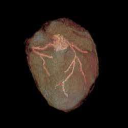
With 3D Heart, coronary arteries can be visualized even at high heart rates (HR-90 bpm, 6-year-old patient with post-AV canal repair).
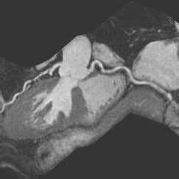
Curved reformat of left and right coronaries.
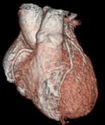
Image courtesy of Funabashi Municipal Medical Center, Japan
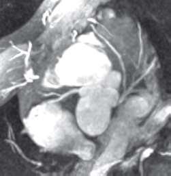
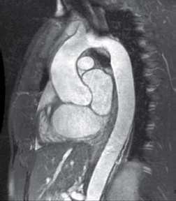
Non Contrast MRA Using 3D Heart
TRICKS
Perform simple, no-miss MRA high in spatial and temporal resolution.
An excellent imaging technique for challenging, contrast-enhanced MRA where timing is critical, TRICKS enables high-resolution, time-resolved vascular imaging without the need for timing or trade-offs between detail and speed. Providing both vessel detail and dynamic information, TRICKS is a very feasible, non-invasive technique for assessing vascular disease — without the use of ionizing radiation.
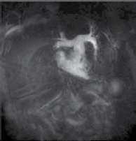
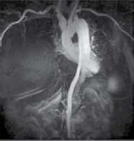
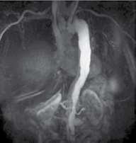
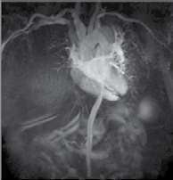
TRICKS showing dynamic filling in an aortic dissection.
High-definition Phase Contrast
Quantify blood flow noninvasively.
Thanks to high-definition Phase Contrast imaging, GE Cardiac MR makes short work of all aspects of blood-flow assessment. Complete measurements quantify blood flow and evaluate flow volumes and velocities — all non-invasively.
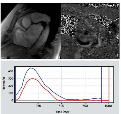
These MR images demonstrate aortic insufficiency. ReportCARD 4.0 quantifies and charts severe AI.
PFO
Detect PFO's with ease.
A leading cause of stroke in patients under age 55, patent foramen ovale (PFO) can be accurately diagnosed with GE Cardiac MR, comfortably and non-invasively. A non-gated IR-FGRE pulse sequence clearly detects PFOs, while ReportCARD 4.0's image interpretation and shunt identification streamline the entire process.
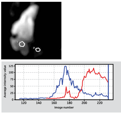
With images generated by our IR-FGRE pulse sequence, detecting PFO's is as easy on your patients as it is for you.
Cardiac MRI Price
| Our Branches | Happy Hours Timings (with prior appointment only) |
|---|---|
|
Alkapuri Branch For Appointment — +91 9227700215 |
Evening — 8:00 PM to 10:00 PM (With Appointment Only)
|
|
Raopura Branch For Appointment — +91 9227510413 |
Morning — 8:00 AM to 10:00 AM (With Appointment Only)
Evening — 8:00 PM to 10:00 PM (With Appointment Only)
|
|
Old Padra Road Branch For Appointment — +91 9227400537 |
Morning — 8:00 AM to 10:00 AM (With Appointment Only)
|
| Study | Price (₹) |
|---|---|
| Cardiac MR | 15,000 |
Why Choose BIC Scans
20+ Years of Trust
Trusted by Doctors
Honest & Transparent Pricing
Present in Vadodara, Bharuch and Surat
More than 2 Lakh Patients Served